S-DMEK in Egypt: First Surgeon, Dr. Ahmed Shaarawy, cornea.clinic Cairo
Dr. Ahmed Shaarawy, founder of cornea.clinic and established corneal transplant surgeon in Cairo, has become the first surgeon in Egypt and the Arab world to perform S-DMEK (S-stamped Descemet Membrane Endothelial Keratoplasty): DMEK using donor tissue that carries a small S-shaped orientation mark. He first presented the technique, with pre-stamped DMEK donor tissue, at the 1st International Egypt and Middle East Congress on Femtosecond Laser Eye Surgeries in Cairo in May 2015. The mark lets the surgeon confirm inside the eye that the graft is the right way up, removing the upside-down graft implantations that were a leading cause of primary graft failure in DMEK[3].
What Is S-DMEK and Why It Matters
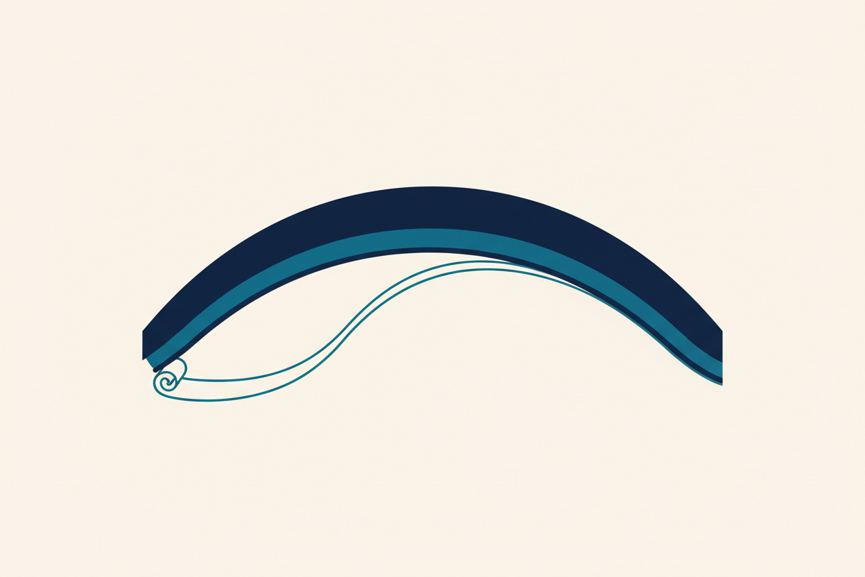
Since it was first described in 2006, Descemet Membrane Endothelial Keratoplasty (DMEK) has become the standard of care for corneal endothelial disease[1][2]. DMEK has one well-known technical hazard, though. The graft is a sheet of Descemet membrane and endothelium only 10 to 15 micrometres thick. It scrolls up on itself, and once unrolled inside the eye it can settle upside down. An inverted graft looks identical to a correctly placed one but cannot pump fluid out of the cornea, so the transplant fails and has to be repeated[3].
S-DMEK, S-stamped DMEK, removes that hazard. Before surgery, the donor Descemet membrane is marked on its stromal side with a tiny S-shaped stamp. Inside the eye a readable S means the graft is the right way up; a mirrored S means it is inverted and must be flipped before the gas bubble fixes it in place. There are no sutures in DMEK; the graft is held against the cornea by an air or gas bubble[3][4].
What does that mean for patients in practice?
- No more upside-down grafts: In the Devers Eye Institute series, primary graft failure caused by inverted grafts was eliminated once S-stamped tissue was introduced[3].
- Fewer repeat operations: A graft that fails because it was inverted has to be replaced. Confirming orientation before the gas bubble is placed avoids that second surgery.
- Less handling of the graft: Orientation is read from the stamp instead of from the direction of the scroll's curl, so the delicate endothelium is manipulated less inside the eye. If the stamp shows the graft is inverted, it can be corrected during the same operation[4].
- Faster visual recovery: Like standard DMEK, vision improves rapidly once the graft adheres and clears.
- Lower rejection rates: DMEK's inherent advantage, minimal foreign surface antigen exposure, remains intact with S-DMEK.
S-DMEK vs. Standard DMEK: The Key Differences
Precision matters here. We use S-DMEK, an advanced corneal transplant technique that uses a specialized, tiny stamp to mark the donor tissue. The mark keeps the tissue correctly oriented during surgery, for the safest and most effective result. The technique was pioneered at the prestigious Devers Eye Institute in Portland, Oregon, the very institution where Dr. Shaarawy completed his specialized cornea fellowship. He trained at the institution where S-DMEK was developed, and performs the technique in Cairo.
Dr. Ahmed Shaarawy: Training Path to Innovation
How does a surgeon become the first in Egypt to perform S-DMEK? For Dr. Shaarawy, the road runs through international fellowship training, and a long-held intent to perform this kind of corneal surgery in the Arab world.
Fellowship at Devers Eye Institute (Oregon, USA, 2014)
In 2014, Dr. Shaarawy completed an advanced fellowship in corneal and anterior segment surgery at the Devers Eye Institute in Portland, Oregon, one of North America's premier centers for keratoplasty, and a place where DMEK techniques are taught, refined, and pushed forward. Under expert mentorship he mastered the full spectrum of DMEK: graft preparation, insertion techniques, troubleshooting, and the management of complications.
That training laid the foundation for moving beyond standard DMEK into specialized variations like S-DMEK. Just as importantly, the Devers years exposed him to emerging innovations in corneal transplantation and to an evidence-based way of thinking about who can, and cannot, be offered surgery.
Return to Cairo: Establishing cornea.clinic
After the fellowship, Dr. Shaarawy came home to Cairo and established cornea.clinic, a practice dedicated entirely to corneal and anterior segment surgery. From day one the clinic has held itself to international standards in patient care, surgical technique, and donor tissue management.
Over the past decade, cornea.clinic has treated hundreds of patients with corneal endothelial disease, building a track record of excellent surgical outcomes. That infrastructure (precise surgical equipment, trained nursing staff, and a structured approach to graft acquisition and preparation) is exactly what made introducing S-DMEK possible.
Founder of ESK-CT: Building Egypt's Corneal Transplant Community
Beyond individual surgical innovation, Dr. Shaarawy has worked to strengthen corneal transplantation as a specialty across Egypt. He founded the Egyptian Society for Keratoplasty and Corneal Transplantation (ESK-CT), an organization dedicated to:
- Advancing surgical standards and training for corneal surgeons in Egypt and the Arab region.
- Promoting evidence-based protocols for donor tissue procurement, evaluation, and transplantation.
- Building collaboration among ophthalmologists and establishing a registry of corneal transplant outcomes.
- Educating patients and the public about corneal disease and the benefits of transplantation.
ESK-CT is part of that same goal: making Egypt a recognized hub for advanced corneal surgery, so Egyptian patients no longer need to travel abroad for specialized treatment.
Why S-DMEK helps in difficult cases
Corneal endothelial disease affects thousands of patients in Egypt and across the Arab world. The causes vary widely:
- Fuchs' endothelial dystrophy (genetic, progressive)
- Post-cataract surgery corneal edema (pseudophakic bullous keratopathy)
- Anterior chamber IOL complications
- Chemical and thermal burns (trauma-related)
- Previous failed corneal grafts
Before S-DMEK, many of these patients faced limited choices:
- Penetrating keratoplasty (PK): A full-thickness graft that requires sutures for 12-24 months, carries higher rejection rates compared to endothelial keratoplasty techniques, and results in significant induced astigmatism.
- DSEK (Descemet Stripping EK): An older partial-thickness technique with slower visual recovery and more corneal scarring than DMEK.
- Best medical management: Hypertonic drops and ointments, which manage symptoms but do not restore clear vision.
S-DMEK gives these patients the clarity and rapid visual recovery of DMEK with the added safety of confirmed graft orientation, so the outcome does not hinge on judging the curl of a transparent scroll through a cloudy cornea.
A few clinical examples where S-DMEK earns its place:
- Fuchs' endothelial dystrophy: The commonest reason for DMEK. The stamp is read through the surgeon's microscope even when the patient's own cornea is swollen and hazy.
- Previous failed graft: A patient whose first graft failed can receive a new DMEK graft, with the stamp confirming orientation in an eye where the view is often poorer than at the first operation.
- Pseudophakic bullous keratopathy: Corneal swelling after earlier cataract surgery. DMEK replaces only the failed endothelial layer through a small incision, and the stamp keeps the procedure predictable.
- Eyes with a limited view: Dense corneal oedema or a shallow anterior chamber make the direction of the graft's curl hard to judge. A stamp that reads correctly or in mirror image is unambiguous.
Dr. Shaarawy's Credentials and Recognition
Dr. Shaarawy's expertise is recognized both internationally and at home:
- American Academy of Ophthalmology (AAO): Active member; participant in continuing medical education and research initiatives.
- American Society of Cataract and Refractive Surgery (ASCRS): Full member; engaged in the global corneal transplant and refractive surgery community.
- Devers Fellowship Training: Recipient of advanced training from one of North America's leading corneal transplant centers.
- Conference Presentations: Presented pre-stamped DMEK donor tissue, a first for Egypt, at the 1st International Egypt and Middle East Congress on Femtosecond Laser Eye Surgeries, Cairo (May 2015); presented a complex DMEK case (brown iris with superficial corneal haze) at the Red Sea Ophthalmology Symposium, RSOS 2017, Jeddah, Saudi Arabia (January 2017).
- Founder and leader of ESK-CT: Establishes and maintains standards for corneal transplantation across Egypt and the Arab region.
The Road Ahead: S-DMEK and the Future of Corneal Surgery in Egypt
The successful introduction of S-DMEK at cornea.clinic matters beyond one surgeon. It shows that advanced surgical innovation is no longer the exclusive domain of major medical centers in the United States, Europe, or Australia. With proper training, infrastructure, and a commitment to excellence, Egyptian surgeons can pioneer these techniques themselves.
As more patients learn what S-DMEK can do, demand is expected to grow. And Dr. Shaarawy's willingness to master this technique, and to teach it, may well encourage other Egyptian and Arab corneal surgeons to expand their own skillsets, raising the standard of care across the region.
ESK-CT gives that ambition a formal home: a mechanism for training the next generation of corneal transplant surgeons in Egypt, so that S-DMEK and other advanced techniques become part of the standard curriculum rather than rare exceptions.
Is S-DMEK Right for You?
Have you been diagnosed with corneal endothelial disease, Fuchs' dystrophy, pseudophakic bullous keratopathy, or keratoconus with endothelial stress, and told your endothelial cell count is "borderline" for standard DMEK? Has a previous graft failed? In either case, S-DMEK may be an excellent option.
The starting point is always a thorough evaluation by a corneal specialist, which includes:
- Specular microscopy to determine endothelial cell count and morphology.
- Anterior segment imaging (slit-lamp, OCT) to assess corneal scarring and anatomy.
- Assessment of previous surgical history and any IOL placement.
- Detailed discussion of expectations, recovery timeline, and potential complications.
Schedule Your Corneal Evaluation Today
If you or someone in your family is struggling with corneal endothelial disease, Dr. Ahmed Shaarawy and the team at cornea.clinic are here to help, with thorough evaluations and access to the latest surgical techniques, S-DMEK included.
cornea.clinic
Jad Tower, 7th Floor
Tahrir Street, Dokki
Cairo, Egypt
Phone: +201111182081
Website: cornea.clinic
Don't let corneal endothelial disease keep chipping away at your vision. Contact us today to schedule a consultation with Dr. Shaarawy.
References
- Melles GR, Ong TS, Ververs B, van der Wees J. Descemet membrane endothelial keratoplasty (DMEK). Cornea. 2006;25(8):987-990. PMID: 17102683
- Deng SX, Lee WB, Hammersmith KM, et al. Descemet Membrane Endothelial Keratoplasty: Safety and Outcomes: A Report by the American Academy of Ophthalmology. Ophthalmology. 2018;125(2):295-310. PMID: 28923499
- Veldman PB, Dye PK, Holiman JD, et al. The S-stamp in Descemet Membrane Endothelial Keratoplasty Safely Eliminates Upside-down Graft Implantation. Ophthalmology. 2016;123(1):161-164. PMID: 26439215
- Veldman PB, Mayko ZM, Straiko MD, Terry MA. Intraoperative S-Stamp Enabled Rescue of 3 Inverted Descemet Membrane Endothelial Keratoplasty Grafts. Cornea. 2017;36(6):661-664. PMID: 28410359
Sources and references
Peer reviewed and professional sources used in the medical review of this article:
- Surgical Technique for Graft Exchange after DALK (Editors' Choice clinical video)Alshaarawy, 2021 · aao.org
- Five-Year Graft Survival and Clinical Outcomes of 500 Consecutive Cases After DMEKBirbal et al., 2020 · Cornea
- Descemet membrane endothelial keratoplasty with a stromal rim (DMEK-S)Studeny et al., 2010 · Br J Ophthalmol